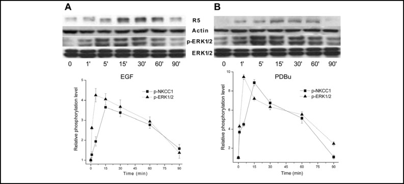
Fig. 3.
Time-dependent changes in ERK1/2 and NKCC1 phosphorylation status induced by EGF and PDBu. HCEC were serum starved for 24 h at 80% to 90% confluence. Panel A shows the effects of exposure to 10 ng/ml EGF for up to 90 min with a representative Western blot analysis of anti-phosphor p-NKCC1 (i.e., R5) and p-ERK1/2 antibody binding. Panel B shows cells exposed to 1 μM PDBu and Western blot analysis performed using the same antibodies as those shown in panel A. Equal loading of proteins was confirmed by reprobing the blots with β-actin.

