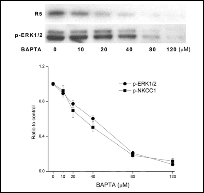
Fig. 6.
Dependence of EGF-induced ERK1/2 and NKCC1 phosphorylation on [Ca2+]i. HCEC were serum starved for 24 h at 80% to 90% confluence. Representative Western blot analysis compares the dose dependent inhibitory effects of exposure to BAPTA on 10 ng/ml EGF-induced p-NKCC1 and p-ERK1/2 formation detected with the anti p-NKCC1 (i.e., R5) and p-ERK1/2 antibodies. HCEC were preincubated for 30 min with each of the indicated BAPTA concentrations prior to exposure to EGF for an additional 5 min. Results were normalized to the increase in p-ERK1/2 formation obtained after 5 min in the absence of BAPTA. Data represent the mean ± SEM of three independent experiments (at concentrations over 40 μM BAPTA, p<0.001 versus untreated control).

