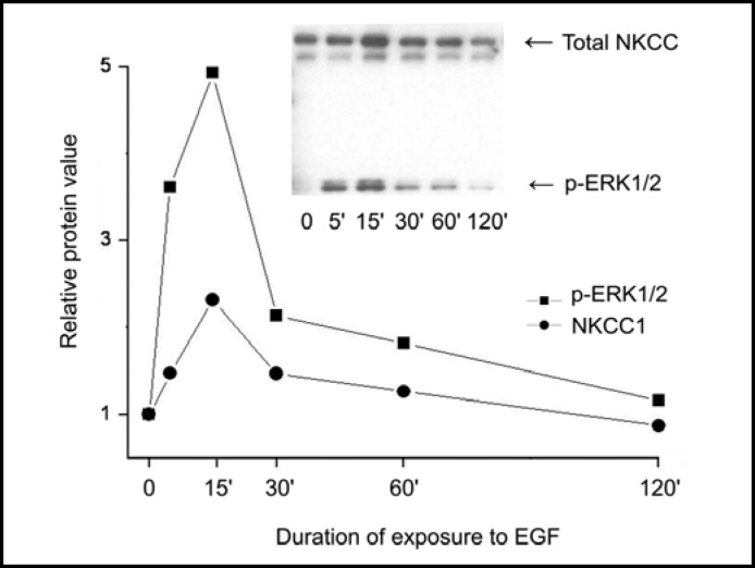
Fig. 7.
Time-dependent changes in p-ERK1/2 association with NKCC. HCEC were serum starved for 24 h at 80% to 90% confluence. Cells were exposed to 10 ng/ml EGF for up to 120 min. Membrane-enriched fractions were obtained following centrifugation. Pellets were probed for relative amounts of total NKCC1 with the anti T4 antibody and p-ERK1/2 antibodies at the indicated times following exposure to EGF. Data represent the mean ± SEM of three independent experiments (after 15 min exposure to EGF, p<0.002 total NKCC versus untreated control; or p<0.001 p-ERK1/2 versus untreated control, respectively). The error bars fall within the range of the indicated symbols for p-ERK1/2 and NKCC1.

