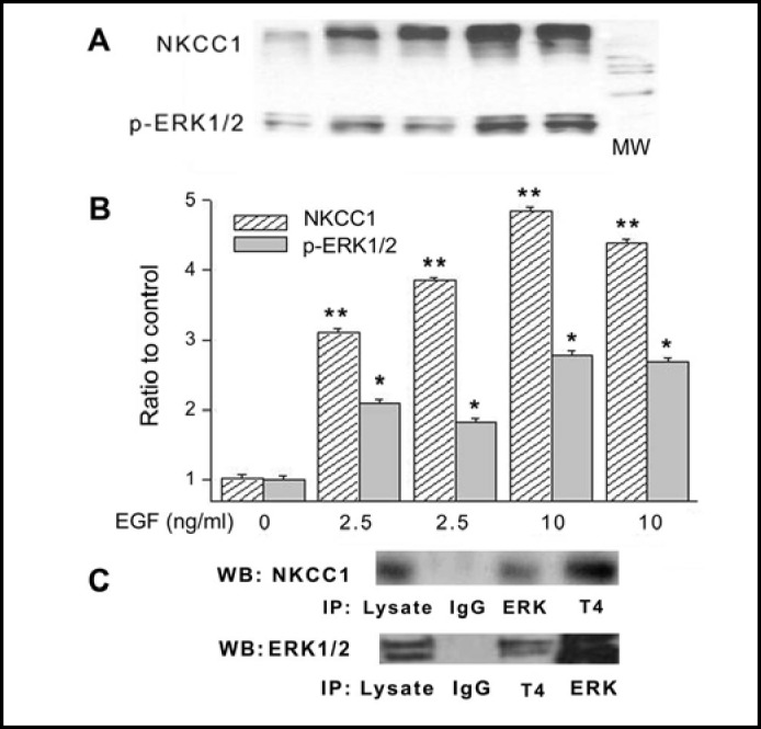
Fig. 8.
Pull-down experiments validate NKCC1-p-ERK1/2 interaction induced by EGF. HCEC were serum starved for 24 h at 80% to 90% confluence. (A) Cells were exposed to either 2.5 or 10 ng/ml EGF for 10 min. Membrane enriched pellets were obtained from different cell lysates and Western blot (WB) probing for either p-ERK1/2 or total NKCC1 association with p-ERK1/2 and T4 antibodies, respectively. (B) Results of experiments performed in duplicate at each concentration normalized to the amounts detected prior to exposure to EGF (Exposure to 2.5 or 10 ng/ml EGF, *p<0.05 p-ERK1/2 versus untreated control; **p<0.001 NKCC1 versus untreated control). (C) Co-localization validation entailed immunoprecipitation (IP) with beads bound to ERK1/2 or NKCC1 followed by probing the precipitate with either anti T4 or ERK1/2 antibodies using Western blot analysis. In the IPs obtained with either the anti T4 antibody or ERK1/2 antibody, there are increases in the amounts of ERK1/2 and NKCC1, respectively. These results validate that exposure to EGF-induces increases in the amounts of p-ERK1/2 associating with NKCC1. Cells were exposed to 10 ng/ml EGF for 10 min. The results shown were obtained from two different experiments each performed in triplicate.

