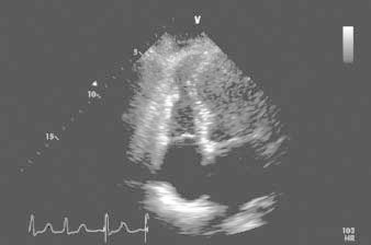
Fig. 2 Transthoracic echocardiogram (4-chamber view), obtained shortly after admission. The associated video shows distal right ventricular free-wall akinesia/dyskinesia, lower septal and apical left ventricular dyskinesia, and cavity dilation.
Real-time motion image is available at www.texasheart.org/journal.
