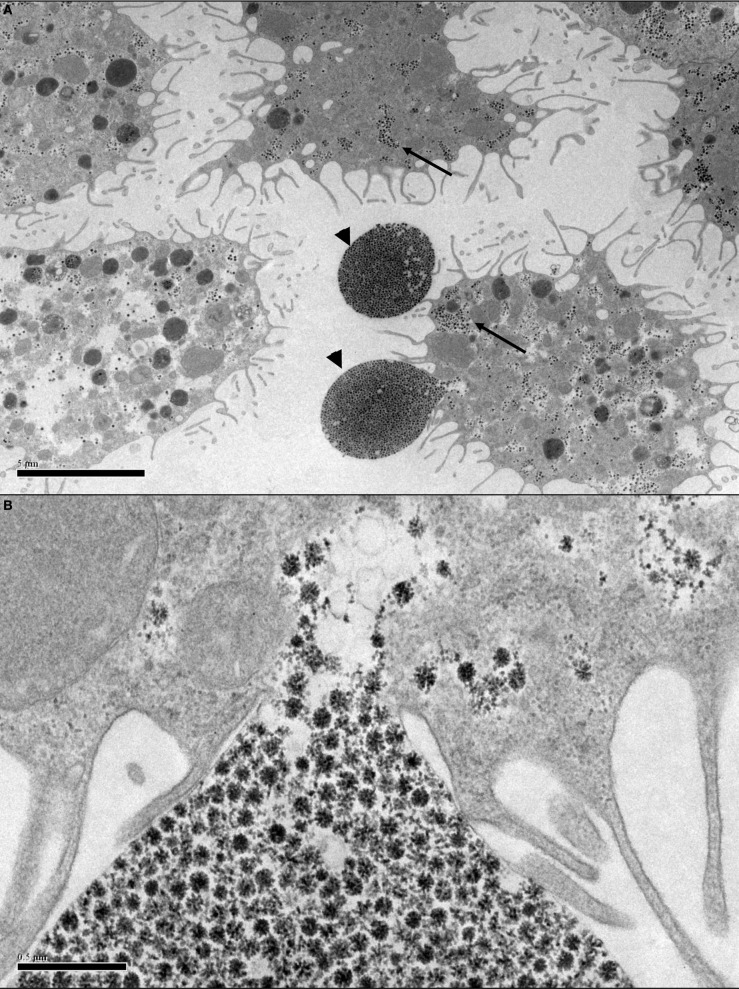
Figure 2.
Transmission electron micrographs of Ornithodoros moubata cell line OME/CTVM25 showing putative endogenous viruses. (A) Cells with “filopodia” extending from the cell surface. Reovirus-like particles (arrows) are abundant in the cytoplasm. The viruses appear to be using the filopodia to form vesicles (arrowheads) carrying a large number of virus particles with diameters of 75–80 nm that may be budding from the cells into the supernatant medium. Scale bar 5.0 μm. (B) Closer view of (A) showing the site of attachment of the virus vesicle to the cell membrane. Scale bar 0.5 μm.

