
Figure 1. Brain abscesses are typified by CD4+ and NKT cell infiltrates.
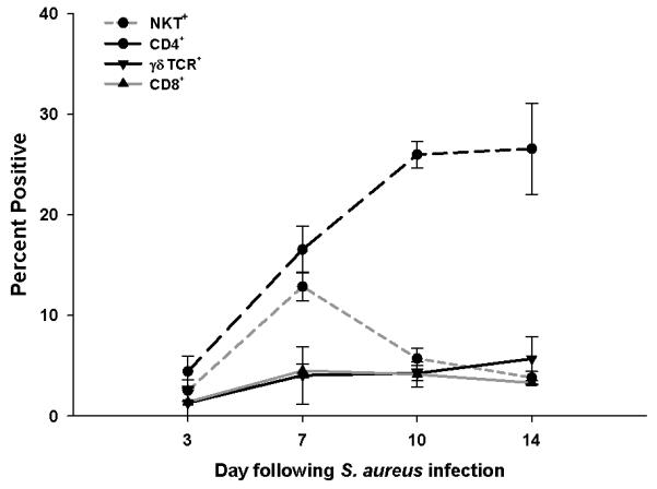
Abscess-associated cells were isolated from C57BL/6 mice at the indicated day post-infection and stained for FACS with NK1.1-FITC, CD4-PECy5, CD8-APC, and γδ TCR-PE. Results represent the percent positive cells of each population combined from a total of five independent experiments (mean ± SEM).