
Figure 6. Loss of TCR αβ cells leads to exaggerated γδ T cell influx in brain abscesses.
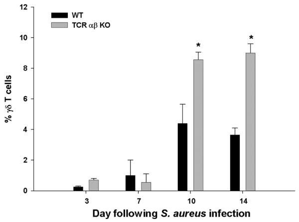
Abscess-associated cells were isolated from TCR αβ KO and WT mice (n= 4-5/group), whereupon the percentages of γδ T cells were identified by FACS. Significant differences between TCR αβ KO versus WT mice are denoted by asterisks (*, p < 0.05). Results represent the mean ± SEM combined from three independent experiments.