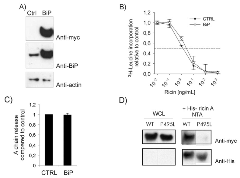
Figure 1.
(A) HEK293 cells were transfected with BiP or an empty vector (ctrl). Lysates were run on SDS-PAGE, transferred to a PVDF membrane and examined for BiP expression using an anti-myc antibody or anti-BiP antibody. Anti-actin was used for loading control. (B) Cells were transfected as indicated and incubated with increasing amounts of ricin in leucine-free medium for 3 h, then washed and incubated with 1 µCi/mL [3H]leucine for 20 min. The incorporation of [3H]leucine was measured and data presented relative to the control (CTRL). This experiment was repeated three times with similar results in duplicates. (C) Cells transfected as indicated were pre-incubated with 0.2 mCi/mL 35SO42- for 3 h and then with ricin sulf-1 for an additional 3 h. The cells were lysed and ricin was immunoprecipitated and subjected to SDS-PAGE under non-reducing conditions. The amount of ricin A-chain reduced from the holotoxin in each sample was quantified and compared to total sulfated ricin in the samples. (D) Cells were transfected with myc-tagged BiP constructs as indicated (wild type, WT or substrate binding mutant, P495L) before lysis and incubated with 1 μg His-tagged ricin A-chain for 1 h. The toxin was pulled down using a Ni-NTA column. The beads were separated in a SDS-PAGE and analyzed by Western blot. BiP proteins were detected using anti-myc antibodies and ricin with anti-His antibodies. Whole cell lysates (WCL, left panels) and pull down (+His-ricin A NTA, right panels) are shown.

