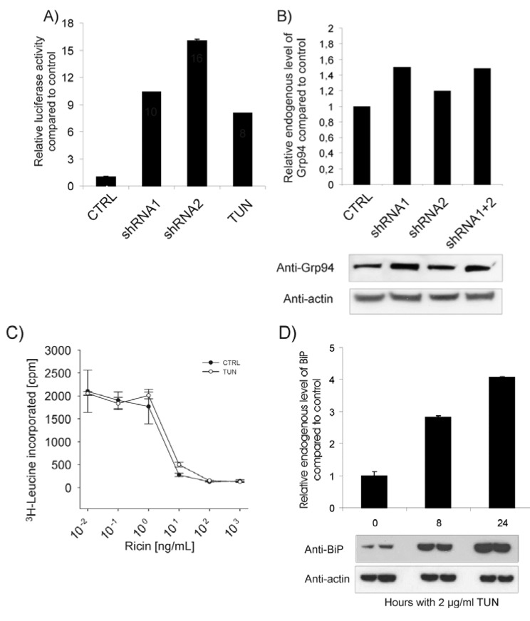
Figure 4.
(A) Cells were transfected with control vector or BiP shRNA constructs together with an ATF6-luciferase reporter construct and Renilla luciferase. 2 μg/mL tunicamycin was used as positive control. Cells were analyzed for luciferase activity 3 days post transfection and the relative activity was normalized to the Renilla luciferase internal control. (B) Lysate from cells transfected with the shRNA constructs were analyzed for the level of Grp94 by Western blot using rat anti-Grp94. The intensity of each band was quantified by ImageQuant 5.0 software. (C) Non transfected cells were incubated with leucine-free medium with or without 2 µg/mL tunicamycin (TUN) for 21 h before different concentrations of ricin were added for subsequently 3 h, then washed in leucine-free medium and incubated with 1 µCi/mL [3H]leucine for 20 min. The amount of radioactive protein was finally measured. (D) Cells were treated with 2 µg/mL tunicamycin (TUN) for 0, 8 and 24 h in parallels before lysis. Lysates were subjected to SDS-PAGE and Western blot analysis. Anti-BiP was used to detect BiP and anti-actin was used as loading control. The intensity of each band was quantified by ImageQuant 5.0 software where endogenous level of BiP at time point 0 was used as the reference level.

