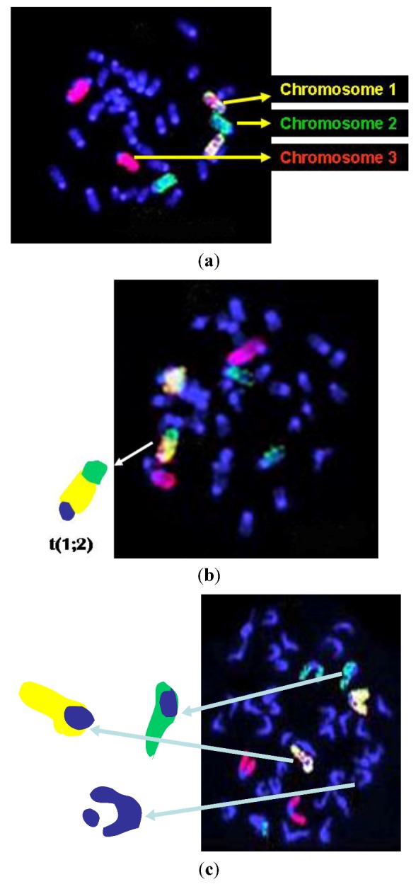
Figure 1.

(a) Representative image of a normal metaphase cell; (b) Representative image of a metaphase with a translocation between mouse chromosomes 1 and 2 (arrow), and (c) Representative image of a metaphase with chromatid breaks on chromosomes 1, 2, and nP.
