
Figure 1.
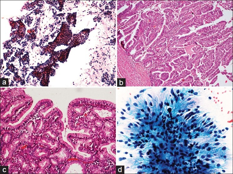
(a) Papillary fragments of tumor cells showing minimal cellular and nuclear pleomorphism, papanicolaou (Pap ×100), (b) intestinal type papillary adenocarcinoma showing true papillae with central fibrovascular cores lined by tall columnar cells and rare goblet cells, H and E, ×100, (c) papillae lined by tall columnar cells along with abundant mucus cells in a case of gastric type of papillary adenocarcinoma, H and E, ×200, (d) columnar tumor cells on cytology, Pap ×400