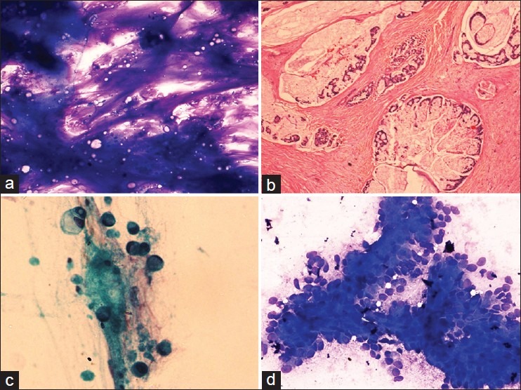
Figure 2.

(a) Tumor cells floating in abundant extracellular mucin, May Grunwald Giemsa (MGG) ×100, (b) mucinous carcinoma showing clusters of tumor cells lying in pools of mucin, H and E, ×100, (c) singly lying tumor cells with abundant intracellular mucin pushing the nucleus to the periphery signifying signet ring appearance, papanicolaou ×400, (d) tumor cells showing moderate nuclear pleomorphism and arranged in cohesive fragments, MGG ×200
