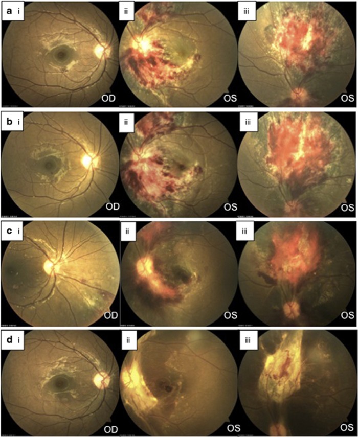
Figure 1.
(a) Fundus appearance on presentation, showing an extensive area of haemorrhagic retinitis inferior to and involving the optic disc associated with gross macula oedema in the left eye (ii), and a second area superiorly (iii), consistent with CMV retinitis. (b) Fundus appearance after 5 days of systemic forscarnet therapy, with worsening of retinitis. (c) After 2 weeks, showing partial resolution of disease in the left eye (ii, iii) with combined systemic and intravitreal treatment, but new lesions in the right eye, both in the macula and inferonasal retina (i). (d) Fundus appearance at 5 weeks after bilateral intravitreal therapy, showing inactive disease with extensive scarring in the left eye.

