
Figure 2.
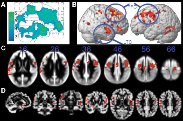
Structural MRI studies showing positive relationships with brain volume and fitness. (A) Gray matter regions, including prefrontal cortex and parietal regions, showing fitness-related preservation in older adults. From Colcombe et al. (2003), Figure 1, p. 178. Adapted with permission. (B) Regions showing increased brain volume in older adults who walked more than 72 blocks per week. From Erickson et al. (2010), Figure 2B, p. 1419. Adapted with permission. (C) Gray matter regions, including bilateral prefrontal cortex, showing a positive relationship with fitness (after controlling for age, gender, and education) in older adults. The blue numbers represent MNI coordinates in the axial (z) plane. From Weinstein et al. (2012), Figure 1A, p. 815. Adapted with permission. (D) Brain regions showing a positive relationship with fitness (after controlling for age, education, and gender) in older adults. From Gordon et al. (2008), Figure 3A, p. 835. Adapted with permission. LTC, lateral temporal cortex; PFC, prefrontal cortex.