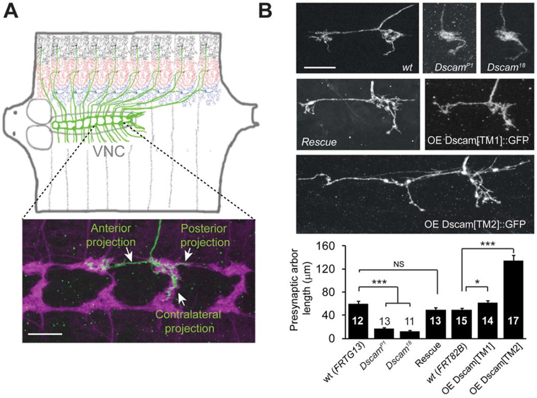
Figure 1. Dscam Instructs Presynaptic Arbor Growth.
(A) Top: Schematic representation of Drosophila larval C4da neurons. In each hemi-segment, Three C4da neurons, ddaC (black), v’ada (red), and vdaB (blue), elaborate dendrites on the body wall and send axons (green) to the VNC to form a ladder-like structure. Bottom: the presynaptic arbor of a single ddaC neuron (green). The flip-out technique was used to express the membrane GFP marker mCD8 ∷ GFP. The presynaptic terminals of C4da neurons, which express another membrane marker, mouse CD2 (magenta), collectively form a ladder-like structure. (B) Representative images and quantification of the presynaptic arbors of single C4da neurons that are wild-type (wt), null mutants of Dscam (DscamP1 or Dscam18), null mutants rescued by one copy of a transgene harboring the Dscam genomic DNA (Rescue), overexpressing the dendritic (OE Dscam[TM1] ∷ GFP) or overexpressing the axonal (OE Dscam[TM2] ∷ GFP) isoform. The MARCM technique was used in these experiments, and the arbors of single ddaC neurons are shown. Sample numbers are indicated in each bar. Scale bars: 10 mm. Also see Figure S1.

