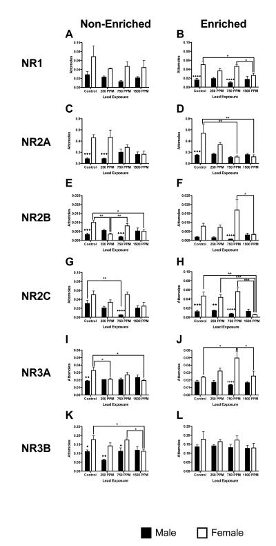
Figure 3.
Effects of perinatal lead-exposure on NMDA receptor subtype gene expression profiles in the hippocampus of male and female rats housed in either a non-enriched or enriched environment. Quantitative PCR analysis of mRNA expression levels for five NMDA receptor subtypes: Nr1 (A, B), Nr2a (C, D), Nr2b (E, F), Nr3a (G, H) and Nr3b (I, J) in the hippocampus of control and lead-exposed animals. Significant sex, environment and dose specific effects were observed. Data are mean number of attomoles of mRNA ± S.E.M. * p<0.05, ** p < 0.01, and *** p<0.001 within sex analysis. ◆ p<0.05, ◆◆ p < 0.01, ◆◆◆ p<0.001 and ◆◆◆◆ p<0.0001 across sex analysis.

