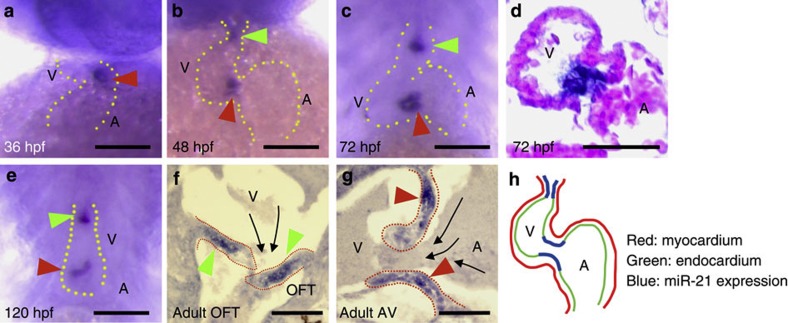
Figure 1. Expression patterns of miR-21.
(a) Soon after initiation of blood flow (36 hpf), miR-21 began to be expressed in a subset of the endocardial cells in the AV canal (red arrowhead). A, atrium; V, ventricle. (b) At 48 hpf, endocardial expression in the OFT became evident (green arrowhead). Expression at the AV canal was arranged around the canal lumen (red arrowhead). (c,d) The same expression patterns were maintained at 72 hpf (c) and 120 hpf (e). (d) In sections, miR-21 expression was detected only in the endocardial cells, not in the myocardial layer. (f,g) Expression of miR-21 was detected in the valves of adult fish at the OFT (green arrowheads in f) and the AV canal (red arrowheads in g). Dotted red lines indicate contours of the valves. Black arrows indicate the direction of blood flow. (h) A schematic representation of miR-21 expression only in specific regions of the endocardium of the OFT and the AV canal (blue lines). Scale bars, 50 μm.

