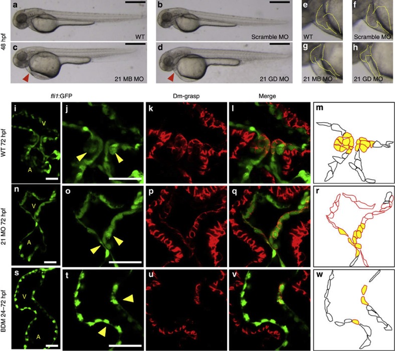
Figure 4. A valveless miR-21 morphant.
(a–d) Blood flow of the miR-21 morphants stagnated with pooling of blood cells on the yolk at 48 hpf (red arrowhead in c,d) (MB, multiblocking MO; GD, Guide Dicer MO). This stagnation was neither present in the WT (a) nor embryos injected with a scrambled MO (b). Higher magnification views are shown in e–h, with heart contours indicated by dotted lines. (i–m) At 72 hpf, thick layers of the GFP-positive endocardial cells were protruded into the AV canal (i), with cuboidal thickening cells (yellow arrowheads in j). Cell shapes are visualized by Dm-grasp staining (k,l). Dm-grasp- and GFP-positive cells are cuboidal, whereas Dm-grasp-negative and GFP-positive cells are squamous (l). (m) A schematic representation of the endocardial cells, with cells in the valve-forming area shown in yellow, Dm-grasp-positive cells in red lines, negative cells in black lines, respectively. A, atrium, V, ventricle. (n–r) In the miR-21 morphants, only a thin layer of endocardial cells was evident with neither thickening nor protrusion into the lumen. (s–w) When heartbeat was stopped from 24 to 72 hpf by BDM, neither thickening nor protrusion of the endocardium was initiated. The number of the Dm-grasp-positive endocardial cells was reduced. Scale bars, 500 μm (a–d), 50 μm (i,j,n,o,s,t).

