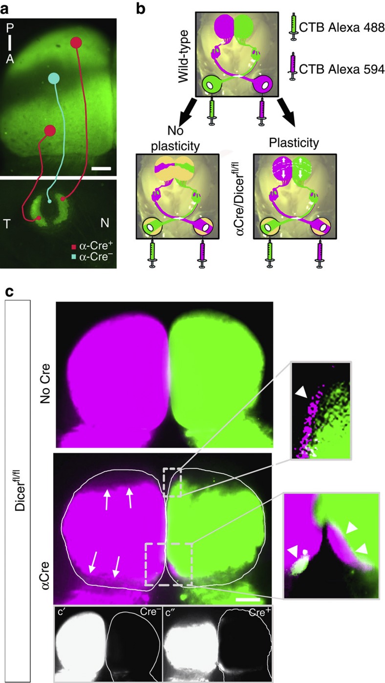
Figure 2. Retinal degeneration results in an extension of the remaining retinocollicular projections.
(a) Dorsal view of the SC (upper panel) and retina (lower panel) at P8 of a αCre+;Dicer+/fl;R26R-EYFP mouse illustrating the Cre-positive areas (green) in the retina and their termination areas in the SC. Nasal (N) and temporal (T) retinal areas map onto posterior (P) and anterior (A) SC, respectively (red lines), whereas the central retina (Cre-negative) maps onto the central SC (blue line). (b) Schematic describing possible outcomes of mapping behaviour of axons originating in remaining central retina, after degeneration of nasal and temporal areas. Without any plasticity (left panel), the RGC axons should map according to their topographic labels and the labelled SC area should thus be confined to the central area, similar to the Cre-negative area shown in (a). If plasticity is involved (right panel) an extension of the labelled area towards anterior and posterior SC is expected. (c) Dorsal view of the SC at P8 of a wild-type (upper panel) and α-Del mouse (lower panel) after full eye fills with CTB-Alexa-488 (green) and -594 (magenta). In wild-type mice, both colliculi are filled completely with axon terminations from the contralaleral eye. In contrast, mice with nasal and temporal degenerations exhibit an incomplete fill of the SC by the projections from contralateral RGCs, leaving some areas at the anterior and posterior end empty (arrows). In addition, small zones occupied by RGC axons originating from the ipsilateral retina are visible in each anterior and posterior SC (arrowheads, boxed areas and higher magnifications). Single channel images (Alexa-594) are shown in c′ and c′′ for increased contrast and signal detection. A–P, anterior–posterior; N–T nasal–temporal. Scale bar, 200 μm.

