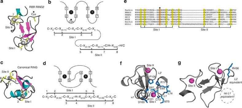
Figure 2. RING2 domain of parkin and other RBR E3 ligases do not recruit E2 enzymes.
Parkin RING2 defines a novel structure common to RBR ubiquitin ligases that are unable to recruit E2 enzymes. The structures and schematic representations of (a,b) RBR RING2 (parkin) and (c,d) RING (TRAF6, PDB 3HCS) domains showing differences in Zn2+ coordination and consensus sequences. (e) Alignment of the RING2 domains of RBR ligases showing residues predicted to coordinate Zn2+ (yellow) based on the parkin RING2 structure and the proposed catalytic cysteine conserved in all sequences (orange). (f) Key residues and elements used for E2 recruitment by the canonical RING E3 ligase TRAF6 are found in two loops (L1, L2) formed through Zn2+ coordination that are absent in (g) parkin RING2. The structures are oriented using the loops involved in Zn2+ coordination for TRAF6 site II and parkin RING2 site I, which exhibit the closest structural similarity.

