Figure 1.

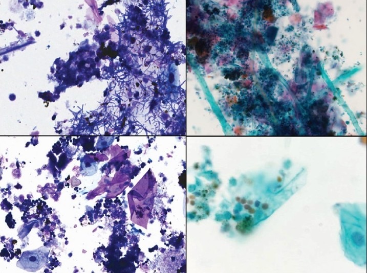
Pleural fluid showing benign squamous cells, debris, bacterial colonies, and fungal colonies consistent with candida. Diff Quik stain, ×400 (left), and Papanicolaou stain, ×200 (right upper) and ×400 (right lower)

Pleural fluid showing benign squamous cells, debris, bacterial colonies, and fungal colonies consistent with candida. Diff Quik stain, ×400 (left), and Papanicolaou stain, ×200 (right upper) and ×400 (right lower)