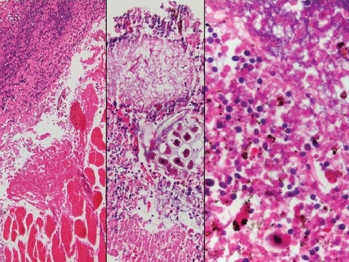
Figure 3.

The surgical specimen of esophageal repair and decortications showing inflammatory exudate involving skeletal muscle (left) and containing vegetable materials (middle), and benign squamous cells bacterial and fungal colonies (right). H and E stain, ×100 (left); ×200 (middle), and ×400 (right)
