Fig. 6.

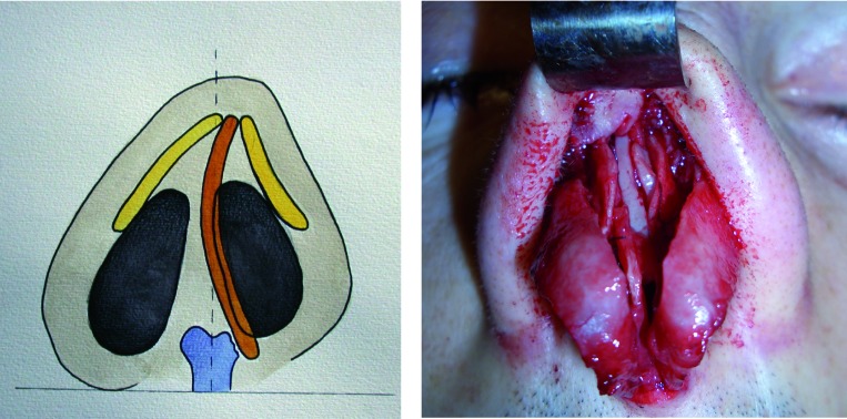
A. Schematic diagram showing the depression of the upper lateral cartilage on the concave side. B. Intraoperative photograph showing lateralization of the left upper lateral cartilage obtained with a septal crossbar graft.

A. Schematic diagram showing the depression of the upper lateral cartilage on the concave side. B. Intraoperative photograph showing lateralization of the left upper lateral cartilage obtained with a septal crossbar graft.