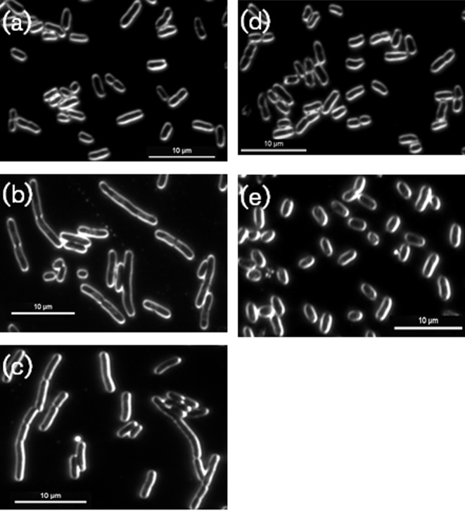
Fig. 2.

Complementation of cell-division defect of E. coli MG1655 hupAB double mutant with PG0121 HU. E. coli strains were grown to mid-exponential growth phase in LB broth and then imaged at 100× magnification with dark field microscopy. (a) E. coli MG1655 parent strain. (b) SG 790; E. coli MG1655 hupAB double mutant. (c) SG 886; E. coli MG1655 hupAB double mutant + empty pTrcHis2A. (d) SG 903; E. coli MG1655 hupAB double mutant + pTrcHis2A expressing E. coli HU-β coding region. (e) SG 918; E. coli MG1655 hupAB double mutant + pTrcHis2C expressing PG0121 HU coding region. The images shown are representative of three independent experiments. Bars, 10 µm.
