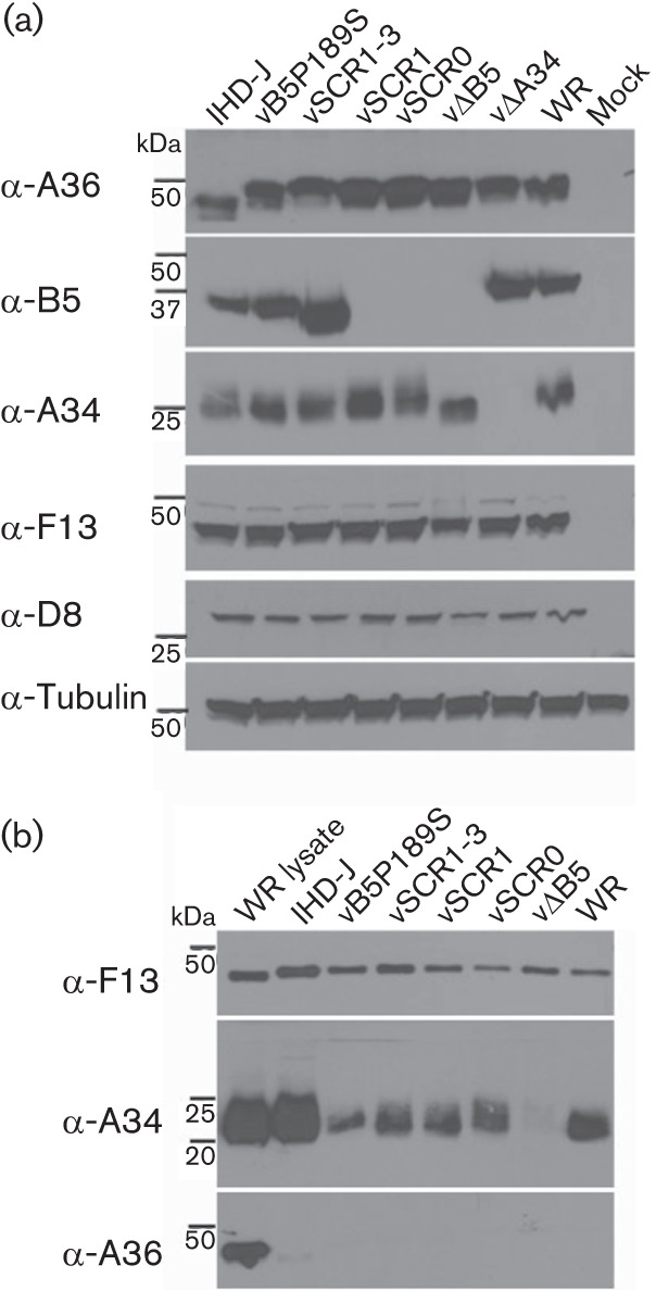
Fig. 6.

Incorporation of A34 into EEV. (a) Expression of IEV/EEV proteins in cell lysates. HeLa cells were infected at 2 p.f.u. per cell with the indicated viruses for 24 h. Protein lysates were prepared and analysed by immunoblotting using antibodies raised against F13, A36, B5 and A34. An anti-tubulin mAb was included as loading control. Note that B5 was not detected in cells infected with vSCR0 and vSCR1 because the rat mAB 19C2 recognizes B5 SCR2 (Law & Smith, 2001). (b) Incorporation of A34 into EEV. RK13 cells were infected at 3 p.f.u. per cell with the indicated viruses for 16 h. EEV were collected, lysed and analysed by immunoblotting.
