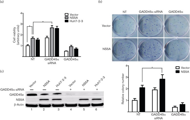
Fig. 4.
Role of GADD45α in HCV NS5A-induced cell proliferation. (a) Stable vector-Huh7, NS5A-Huh7 and Huh7-2-3 cells were seeded into a 96-well plate 24 h before transfection with GADD45α siRNA (50 nM) or GADD45α plasmid (0.5 µg), as indicated. The cells were incubated for 72 h before the cell viability assay. Results are shown as means±sem of three independent experiments. *P<0.05. (b) The effect of GADD45α on cellular growth was further confirmed by a colony formation assay. The upper panel shows representative images of colony formation in stable vector-Huh7 and NS5A-Huh7 cells transfected with GADD45α plasmid or GADD45α siRNAs. Quantitative analysis of colony numbers is shown in the lower panel (means±sem of three independent experiments). (c) Western blotting confirmed that the GADD45α protein was knocked down by GADD45α siRNAs. The NS5A protein was detected in stable NS5A-Huh7 and Huh7-2-3 cells.

