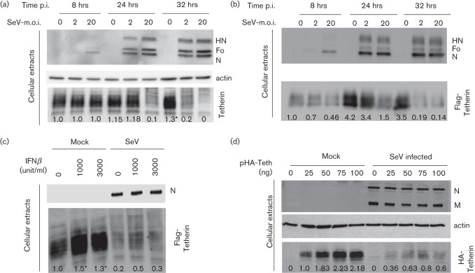
Fig. 4.
Tetherin degradation as a function of infection loads. (a) HeLa cells and (b) 293T cells expressing Flag-tetherin were mock-infected or infected with SeV (m.o.i. = 2 and 20) for 8, 24 and 32 h. Cell lysates were collected and protein levels were analysed by Western blot. (c) HeLa cells pre-treated during 16 h with increasing amounts of interferon β (0, 1000 or 3000 units ml−1) were mock-infected or infected with rSeV (m.o.i. = 20). At 24 h p.i., cell extracts and viral supernatants were analysed by Western blot. (d) HeLa cells were transfected with increasing amounts of a plasmid expressing HA-tagged tetherin (pHA-Teth), 0–100 ng) for 48 h and then infected with SeV (m.o.i. = 20). Cell extracts and viral supernatants were harvested at 24 h p.i. and the expression of the indicated proteins was analysed by Western blot. *, Partially saturated signals (see Methods).

