Fig. 4.
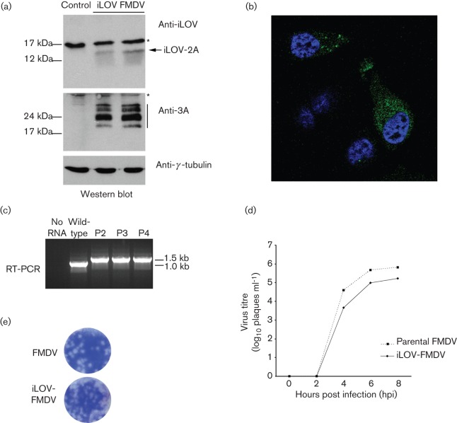
iLOV-FMDV is viable and stable. (a) Western blot analysis of whole-cell lysates prepared from goat epithelium cells infected with iLOV-FMDV. Samples were probed for the presence of iLOV, the non-structural FMDV 3A protein (and 3A-B precursors, black bar) and γ-tubulin (loading control). Asterisks indicate non-specific bands. (b) Fluorescence microscopy of goat epithelium cells infected with iLOV-FMDV. Nuclei are stained blue (DAPI). (c) RT-PCR analysis of iLOV-FMDV genomes prepared from P2, P3 and P4 virus stocks. Genome derived from the parental wild-type virus was also analysed as a control. (d) Growth analysis comparison of parental wild-type FMDV and iLOV-FMDV. Goat epithelium cells were infected with the respective virus (0.1 m.o.i.) and samples analysed at 0, 2, 4, 6 and 8 h post-infection. Similar results were obtained from three individual experiments. (e) Plaque morphology of goat epithelium cells infected with parental wild-type FMDV or iLOV-FMDV.

