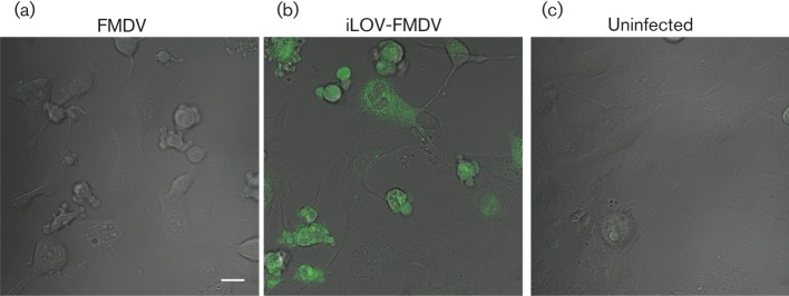
Fig. 6.

Detection of iLOV-FMDV-infected cells by live-cell imaging. (a–c) Still images from live-cell imaging experiments during which goat epithelium cells were infected at an m.o.i. of 2 with either parental-FMDV (a) or iLOV-FMDV (b) or mock infected (c). Cells imaged by differential interference contrast optics are shown. iLOV-expressing cells (green) are clearly visible in (b). Bar, 20 µm.
