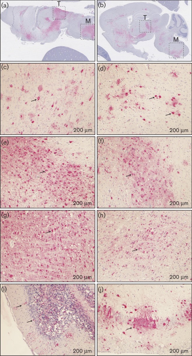
Fig. 6.

RVFV antigen distribution in mouse brain after vaccination. Brain tissues derived from euthanized moribund inbred mice after vaccination with MP-12 (a, c, e and g) or rMP12-TOSNSs (b, d, f, h, i and j) were subjected to immunohistochemistry using anti-RVFV N antibody. Signal specific to RVFV N antigen was developed as red, while counterstain with haematoxylin is shown as blue. (a and b) Overall antigen distributions in brain tissue are shown as red signals. T, Thalamus, M, medulla. Antigen distribution in cerebral cortex (c and d), thalamus (e and f), medulla (g and h), cerebellum (i) and hippocampus (j) are shown. Arrows indicate cells with RVFV N antigens. Bars, 200 μm.
