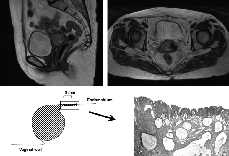
Fig. 5.
T2-weighted MRI images of a 59-year-old patient demonstrated a hyperintense tumor in the right wall of the cervix with a maximal size and length of 31 and 24 mm, respectively. Although there was a part where low signal intense stroma looked to be interrupted, no clear evidence or parametrial invasion is indicated on MRI. On microscopy, the maximal tumor length was measured as 30 mm including an 8-mm lesion of in situ endometrial involvement (hematoxylin and eosin, ×10 magnification).

