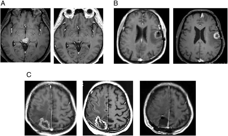
Fig. 2.
Gd-enhanced T1-weighted MR images. (A) Breast cancer brain metastases in a 55-year-old female. A tectum tumor treated with a marginal dose of 30 Gy in three fractions at 60% isodose (left). A significant tumor response and no adverse imaging effects found 11 months after three-fraction CyberKnife radiotherapy (right). (B) Lung cancer brain metastasis in a 64-year-old male. A tumor in the speech area with perifocal edema treated with a marginal dose of 25 Gy in three fractions at 60% isodose (left). A tumor response found 1 year after three-fraction radiotherapy (right). (C) Breast cancer brain metastasis in a 37-year-old female. A tumor in the sensory area with perifocal edema treated with a marginal dose of 30 Gy in three fractions at a 59% isodose (left). A tumor response was found 11 months after treatment. However, both clinical and radiological deterioration were noted (center) and surgical resection was required after osmo-steroid therapy. The surgical specimen confirmed as radiation necrosis and the hemiparesis improved after surgery (right).

