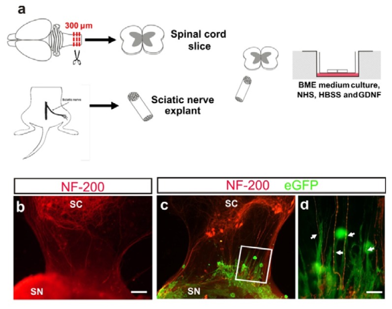
Figure 5.
(a) Representation of spinal cord and sciatic nerve co-culture (see material and methods for details). (b) Fluorescence photomicrograph illustrating an example of organotypic co-cultures of spinal cord (SC) and peripheral nerve (SN) in absence of M-OECs. After seven days in vitro (DIV), axons from motor neurons (red) reach and innervate the sciatic nerve explant. (c,d) Example of the morphology and behavior of M-OECs after transplantation in the organotypic slices. Numerous eGFP-positive cells can be seen, and some of them oriented parallel to the motor neuron axons (arrows in d). Scale bars: b, c = 150 μm, d = 50 μm.

