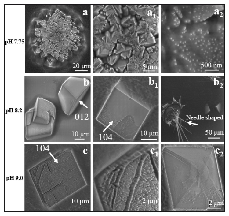
Figure 5.
SEM image of CaCO3 crystal assembly in presence of AS8: (a) polycrystalline spherical assembly at pH 7.75; (b–b2) rhombohedral crystal of calcite in two different orientations: (b) (012) and (b1) (104); (b2) needle shaped peptide crystals at pH 8.2; (c, c1) calcite crystal with distorted face and corners observed at pH 9.0. The SEM images were taken at 10 keV (c, c1: 5 keV), spot size 3 (1.7 nm; c, c1: 2.1 nm). Magnifications: (a) 2000×; (a1) 10000×; (a2) 100000×; (b) 4000×; (b1) 5000×; (b2) 500×; (c) 5000×.

