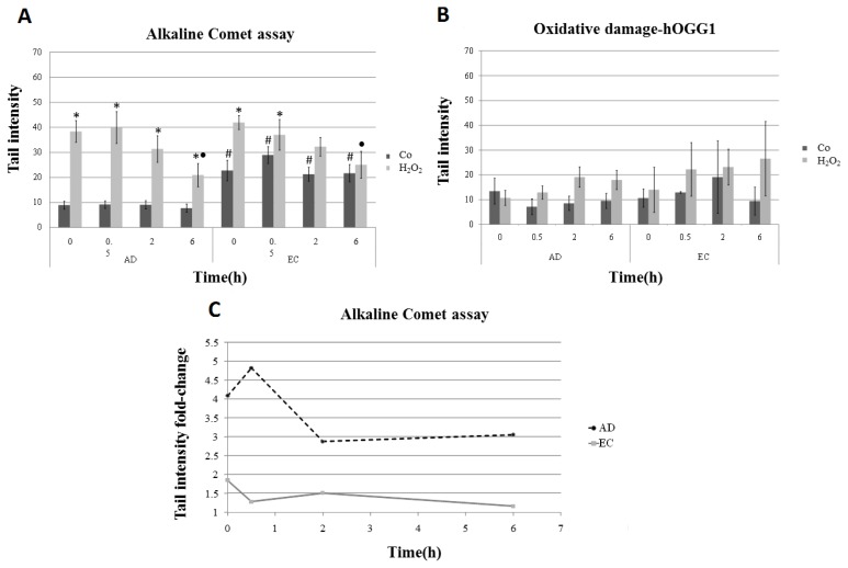
Figure 1.
DNA damage measured as tail intensity (% of DNA in the tail) in the comet assay. Lymphocyte cultures from AD and EC groups of individuals were submitted to treatment with hydrogen peroxide (H2O2) for 1 h and harvested at different recovery times (0, 0.5, 2 and 6 h) after treatment. (A) DNA damage analyzed by alkaline comet assay; (B) estimated net amount of oxidative damage: each value of tail intensity obtained in the conventional comet assay (without hOGG1 enzyme) was subtracted from that observed in the assay performed with the addition of hOGG1; (C) scatterplot showing the extent of DNA damage presented by treated samples in relation to the control samples; fold-change was calculated using mean values of tail intensity. * Statistically significant difference between each treatment with H2O2, and its respective control (p < 0.05); ● Statistically significant difference between the mean values of tail intensity obtained at 0.5, 2 and 6 h of recovery relative to the corresponding initial time (0 h) (p < 0.05); # Statistically significant difference when the EC sample was compared with the corresponding AD sample (p < 0.05).

