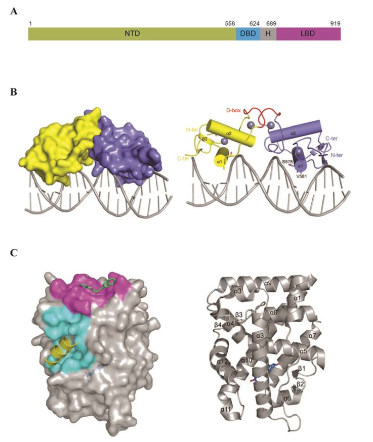
Figure 2.
Domain organization and available structures of the androgen receptor. (A) Scheme of the domain organization of the AR: NTD (N-terminal domain), DBD (DNA binding domain), H (hinge region) and LBD (ligand binding domain). Residue numbers above the scheme delineate the domain boundaries; (B) Surface (left) and cartoon (right) representations of the rat AR-DBD structure. Zinc ions are presented as grey spheres, and the D-box of each DBD monomer is highlighted in red. Residues of the P-box involved in DNA recognition are shown as sticks; (C) Surface (left) and cartoon (right) representations of the human AR-LBD structure: surfaces highlighted in cyan and magenta correspond to AF2 and BF3, respectively. The cartoon representation in yellow corresponds to a coactivator bound to AF2, and the stick representation in green depicts a BF3 small molecule inhibitor. R1881, a synthetic androgen, bound to the androgen binding site, is shown in blue.

