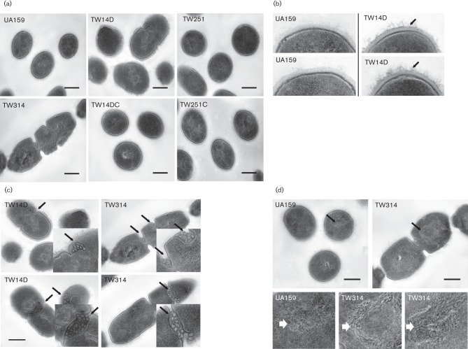
Fig. 4.
TEM analysis of Strep. mutans strains grown in BHI to mid-exponential phase (OD600 0.4). (a) Shows defects in cell division in TW14D and especially, in TW314; (b) highlights rougher, fibrous outer surface (peptidoglycan) in TW14D (indicated by arrows); (c) shows increased presence of mesosomes in TW14D and TW314 (marked by arrows), with inserts showing magnification of the regions indicated); and (d) shows looser nucleoid structure of TW314 (indicated by arrows), as compared to UA159, with magnified images of the selected regions shown below. Images were taken at magnifications of 25K and 60K. Scale bars (all images), 250 nm.

