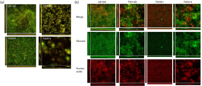
Fig. 5.
Biofilm analysis. Strep. mutans biofilms were grown on hydroxylapatite discs in BM medium supplemented with glucose, sucrose, and glucose plus sucrose for 48 h. Following proper staining using BacLight live/dead fluorescent dye, biofilms were optically dissected using an Olympus laser scanning confocal microscope, and post-acquisition analyses were performed using SLIDEBOOK 5.0 and COMSTAT 2.0. As shown in (a), biofilm formation by TW14D, TW251 and especially TW314 during growth in BMGS was reduced significantly, when compared to the UA159. Relative to UA159, alterations in biofilm structure were also apparent, especially with TW314. (b) To examine glucan production by Strep. mutans, biofilms were treated with Alexa 488-conjugated Concanavalin A and SYTO 59, prior to optical dissections. The green fluorescent glucans (glucans) and red fluorescent cells (nucleic acids) and the merged (merge) images of UA159, TW14D, TW251 and TW314. In comparison, TW251 biofilms had significantly less glucans than the parent strain UA159. Images presented here are representatives composed of xyz, xz and yz images (512×512).

