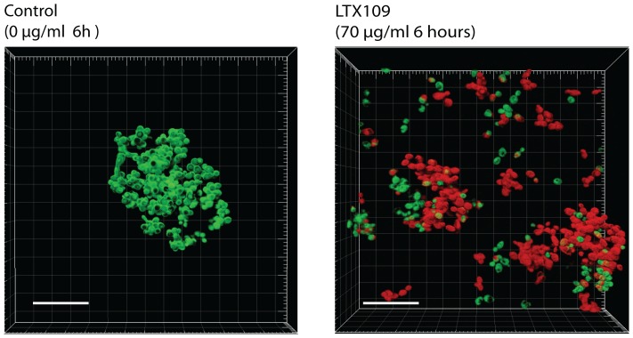
Figure 5. Activity of LTX109 against yeast biofilm.
Confocal Laser Scanning Microscopy of S. cerevisiae (Σ1278b) biofilm. Cells were grown in Lab-Tek™ Chamber Slide™ System; Permanox - (NUNC, Denmark) in 1 ml synthetic complete medium After 12 hours, the cells were exposed to 0 µg/ml LTX109 (control) or 70 µg/ml LTX109 for another 5 hours. The biofilm cells were then stained with Syto9 (green) and propidium iodide (red) LIVE/DEAD stain before confocal laser scanning microscopy. Images are 3D reconstructions of biofilm made from 2 µm thick images in stacks of 20 individual images. CLSM was perform with a Zeiss LSM510 microscope using a 63x/0.95NA a water immersion lens. Life dead staining of biofilm treated with LTX109 was repeated in four independent experiments. White bar is 30 µm.

