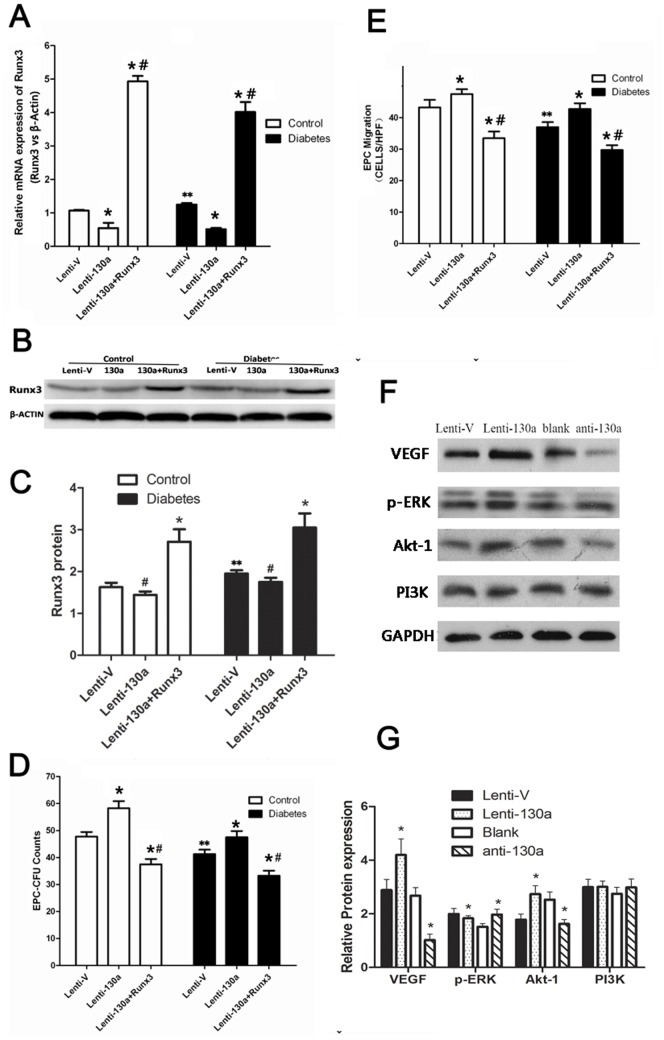
Figure 6. Runx3 mediated miR-130a’s effect on EPC function and miR-130a upregulated VEGF, p-ERK and Akt1.
(A–E) Cells were transfected with both lentiviral miR-130a and lentiviral Runx3 or lentiviral miR-130a alone. (A) mRNA expression of Runx3 by real-time PCR (normalized to β-actin). (B, C) Protein expression of Runx3 by Western blotting. (D, E) Colony formation and migration capacity of EPCs. * P<0.05 vs. respective empty vector groups (Lenti-V). # P<0.05 vs. lenti-130a group. ** P<0.05 between the same processed healthy and diabetic groups (eg. Lenti-130a control groups VS Lenti-130a diabetes groups ) (F, G) EPCs were transfected with a miR-130a inhibitor or a negative control (Blank), or lentiviral miR-130 or an empty vector (Lenti-V). Protein levels of VEGF, p-ERK and Akt1 were measured by Western blotting. *P<0.05 Lenti-130a vs. Lenti-V group or anti-130a vs. scrambled control (blank) group. VEGF: vascular endothelial growth factor; ERK: extracellular signal-regulated kinase; PI3K: phosphatidylinositol 3′-kinase. The presented experiment is a typical result obtained from three separate experiments.

