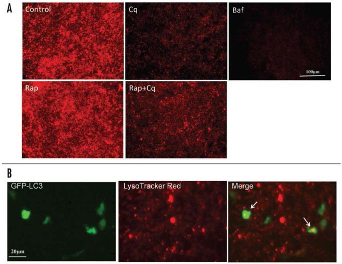
Figure 3.
The effect of chloroquine on lysosomal activity in HL-1 cardiac myocytes. (A) HL-1 cardiac myocytes were incubated with saline (Control), rapamycin (Rap, 5 μM), chloroquine (Cq, 3 μM), rapamycin plus chloroquine (Rap+Cq, 5 μM and 3 μM, respectively), or Bafilomycin A1 (Baf, 50 nM), in serum free media for 2 hr. Following treatment, cells were loaded with 50 nM LysoTracker Red for 5 min in the culture medium and analyzed by fluorescence microscopy. LysoTracker Red assessments were performed in three independent experiments; results presented are representative. (B) Higher magnification image of cells transfected with GFP-LC3 and loaded with LysoTracker Red, showing colocalization of GFP-LC3 and LysoTracker Red in a subset of structures (arrows).

