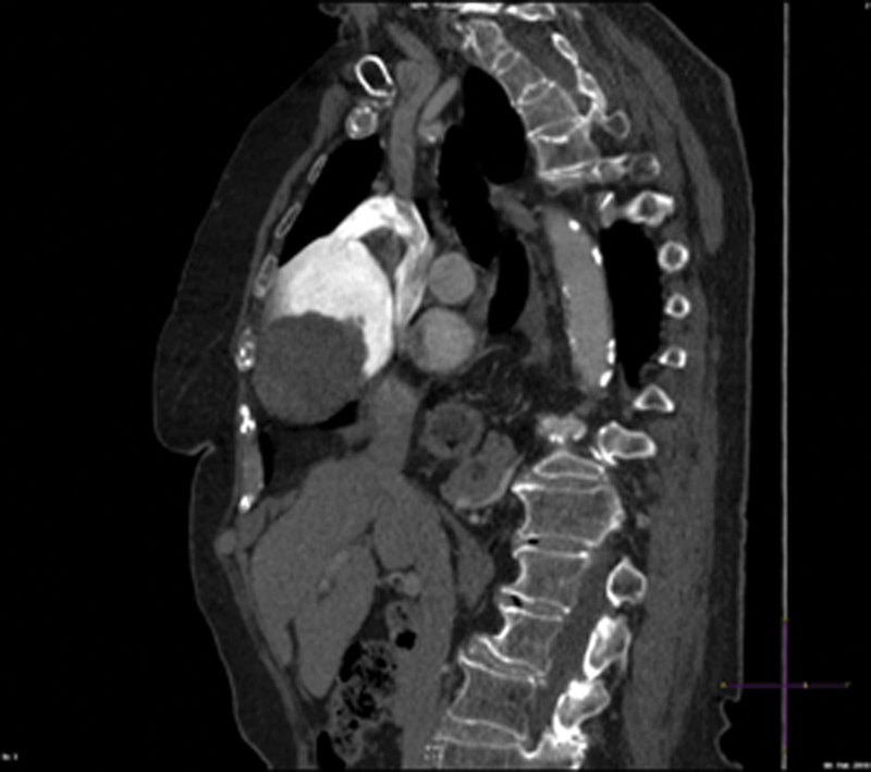
Fig. 5.

Reconstructed multiplanar CT slice in a sagittal oblique plane demonstrates the left innominate vein with contrast and its confluence with the right internal jugular vein as they empty into the superior vena cava. There is a distinct fat plane between the superior vena cava and the left innominate vein aneurysm.
