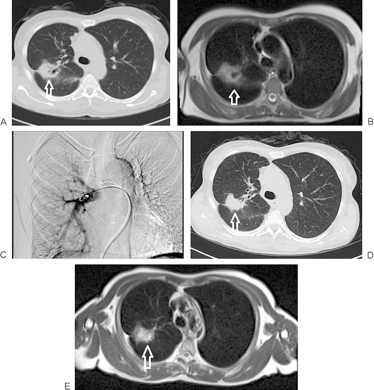
Figure 1.

A 60-year-old woman with primary non-small cell lung carcinoma in the right lung, segment 6, undergoing treatment with transpulmonary chemoembolization. (A) Nonenhanced computed tomography (CT) image of lung before chemoembolization, demonstrating a 41 × 35 mm right lobe lesion (arrow). (B) T2-weighted nonenhanced magnetic resonance imaging (MRI) (2300/90 [TR/TE]) demonstrating the pretreatment tumor extension in the lung (arrow). (C) Angiographic verification of the catheter position in the main right pulmonary artery during the treatment. (D) CT image of the same patient during follow-up at 9 months, demonstrating significant downsizing of the tumor volume (36 × 25 mm) (arrow). (E) T2-weighted nonenhanced MRI of lung on 9-month follow-up, again demonstrating significant shrinkage of the tumor (arrow).
