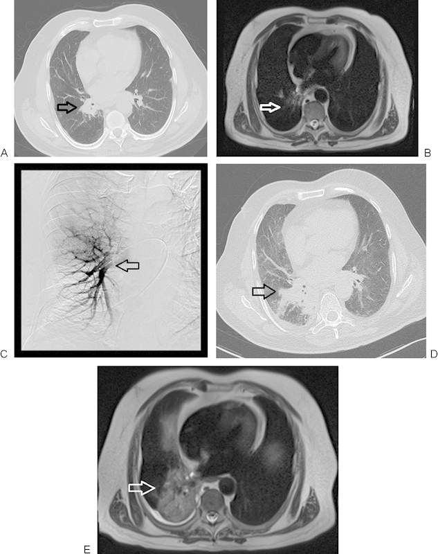
Figure 2.

A 71-year-old man with primary non-small cell lung carcinoma in a right perihilar location, with progression following regional chemoembolization. (A) Nonenhanced computed tomography (CT) image of lung before chemoembolization. The initial lesion measured 39 × 32 mm (arrow). (B) Axial T2-weighted nonenhanced magnetic resonance imaging (2500/90 [TR/TE]) presenting primary lung cancer spreading before chemoembolization (arrow). (C) Transpulmonary chemoembolization with catheter placement in the right perihilar region (arrow). (D) Axial CT follow-up study 4 months after transpulmonary chemoembolization (TPCE) demonstrating tumor progression (59 × 58 mm) (arrow). (E) Axial T2-weighted nonenhanced MRI of the lung, 4 months post-TPCE with further tumor progression (arrow).
