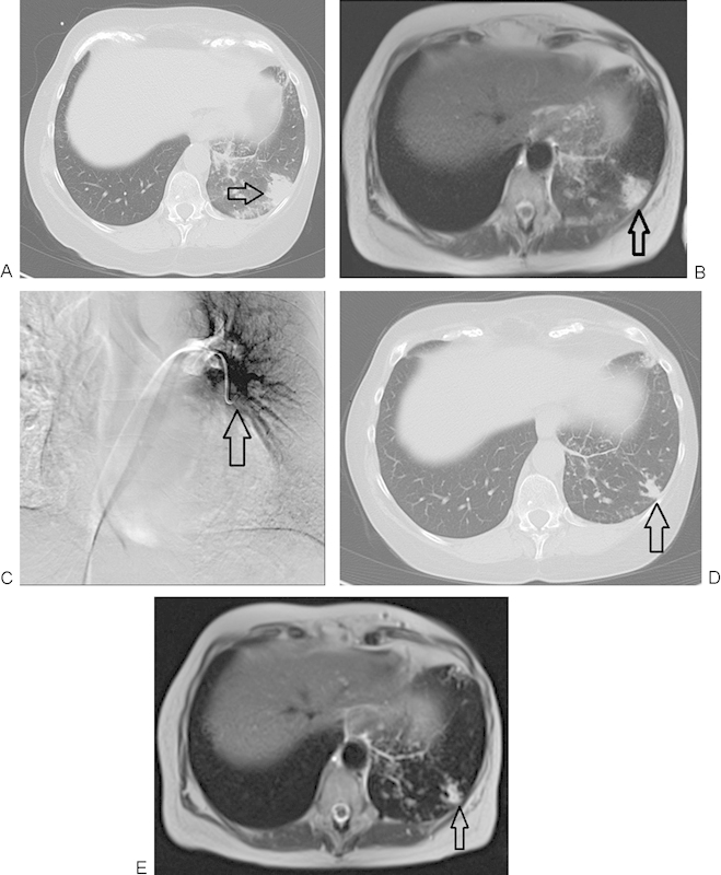
Figure 3.

A 64-year-old woman with breast cancer therapy and resistant lung metastasis. (A) Nonenhanced computed tomography (CT) image of the lung demonstrating metastatic breast cancer before chemoembolization. Lesion initially measured 27 × 26 mm in diameter (arrow). (B) Axial T2-weighted nonenhanced magnetic resonance imaging (MRI) of the lung demonstrating metastasis of lung cancer before chemoembolization (arrow). (C) Transpulmonary chemoembolization (TPCE) with catheter positioned in the middle segment of the left lung (arrow). (D) Axial CT follow-up study 12 months post-TPCE with significant downsizing of the tumor, now measuring 15 × 10 mm (arrow). (E) Axial T2-weighted nonenhanced MRI of the lung in a follow-up study 12 months post-TPCE. Again demonstrated is the significant decrease in lesional diameter (arrow).
