
Figure 1.
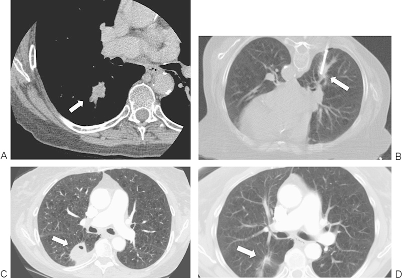
A 74-year-old woman with a non-small cell lung cancer in the right lower lobe referred for radiofrequency ablation (RFA). (A) Axial contrast-enhanced computed tomography (CT) image shows a 2.5-cm mass (arrow). (B) Axial CT image with patient prone during RFA shows a single RFA electrode (arrow) in the center of the mass. (C) Axial contrast-enhanced CT image at 3-month follow-up shows reactionary inflammation and enhancement (seen on other windows) along the adjacent pleura (arrow); this is a normal postablation finding and does not represent residual disease. (D) An axial contrast-enhanced CT image at 31-month follow-up shows near complete involution of the thermal scar (arrow).