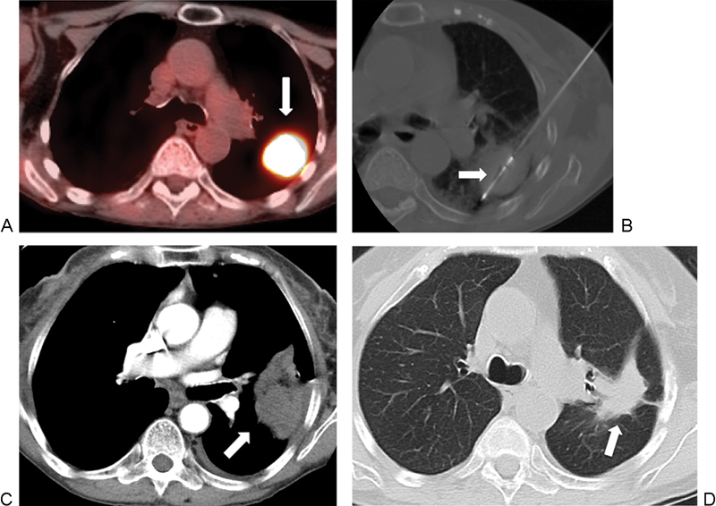
Figure 2.

A 71-year-old woman with metastatic transitional cell carcinoma to the lung, referred for microwave ablation (MWA). (A) Axial positron emission tomography-computed tomography (PET-CT) image shows intense avidity in the left lower lobe, with a mass measuring 3.5 cm (arrow). (B) Axial CT image shows the ablation of the tumor using a 4-cm active tip MW antennae (arrow). (C) Axial contrast-enhanced CT image, mediastinal windows, 15 days after ablation shows lesion consolidation with peripheral and pleural reactive enhancement, which is a normal postprocedure imaging finding. (D) Axial CT 18 months after treatment shows a contracting thermal scar (arrow).
