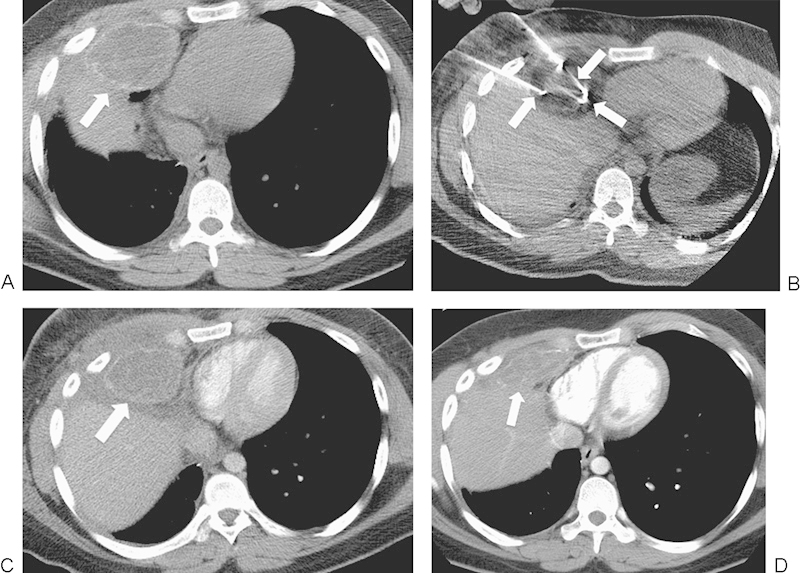
Figure 3.

A 22-year-old man with widely metastatic osteosarcoma presented for palliative cryoablation treatment of an osteosarcoma metastasis in the right middle lobe, invading the chest wall and abutting the right atrium of the heart and the liver. (A) Axial computed tomography (CT) image shows a 6.9-cm mass (arrow). (B) Axial CT image shows three of the six cryoprobes (arrows) in the treated mass. There is a low attenuation ice ball surrounding the lesion. (C) Axial contrast-enhanced CT image 10 days after ablation shows a hypodense response consistent with cell death at the treatment site. Also, mineralization related to the original osteosarcoma is present at the margin of the tumor (arrow) and does not represent residual disease. (D) Axial contrast-enhanced CT image 1 year after ablation shows interval contraction of the ablation zone (arrow).
