
Figure 4.
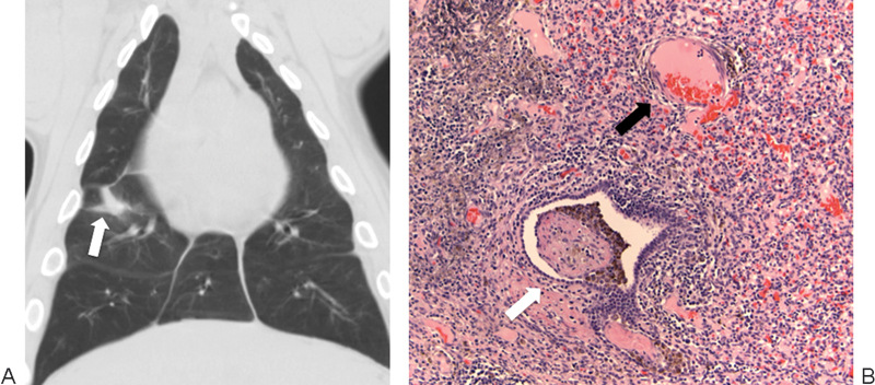
Irreversible electroporation (IRE) in a swine lung model. (A) Coronal reconstruction of a chest computed tomography image 2 weeks after lung IRE shows a fibrotic scar in the right lung (arrow). (B) High-power magnification with hematoxylin and eosin staining of an IRE ablation zone 2 weeks after treatment shows inflammatory infiltrate with intact veins (black arrow) and bronchioles (white arrow).